Debbie update... Well, we're back – and with unexpected news. Good news, mainly.
When we checked in for her appointment, we were surprised by a request to take more X-rays. This request came before we'd even finished the check-in paperwork. Though we'd brought our iPads so we could read while waiting, we never even got a chance to crack them open. The MRI from last week was full of artifacts from the metal in Debbie's knee (we knew this already), so the surgeon wanted X-rays to get a good picture of her knee's current status. These X-rays went quickly and easily, in no small part because of the kind and attentive work of the X-ray tech, a young lady named Brook.
As soon as we were done with the X-rays, we were scooted into the exam room, where we waited for about 10 seconds for the surgeon (Dr. Tom Higginbotham) to come in. We'd not met him before, so we didn't know what to expect, though he was highly recommended to us by two friends in Paradise. Within a few minutes, we knew we had a good 'un – he was cheerful, considerate of Debbie, full of information, and happily answered all our questions.
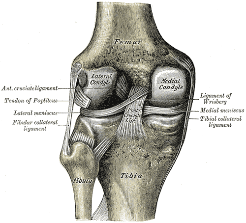
But what he had to say quite surprised us! While he acknowledged that the MRI shows a “slightly ratty” meniscus, he's not at all sure that's a problem. And, more importantly, it's not the worst problem in her knee. He said that the MRI also shows a fracture of her medial condyle (the knobby “knuckle” of the knee, one of two attached to each femur, or upper leg bone). The fracture was just a hairline crack, and the X-rays confirmed that there was no displacement (bones shifting) at all. The treatment for this is very simple: wait. Wait for about six weeks post-injury, or about four weeks from now, before putting any weight on that leg. No surgery required.
The way this fracture was diagnosed was interesting. The MRI clearly shows the edema in her knee. “Edema” is just a fancy word for swelling, in this case in her knee. It was very evident indeed at the time the MRI was taken, and indeed it still is. The MRI also shows that there were two kinds of liquid causing the edema. One was liquid that was mostly water, but the other was a liquid that was mostly fat or oil, and it floated on top of the water (much like separated salad dressing). The water-based liquid is mainly from blood or plasma, but what's the fatty liquid from? Well, it turns out that the presence of such a fatty liquid is a signature of a fracture. It's caused by bone marrow leaking out of the broken bone. It was the presence of that fatty liquid that lead Dr. Higginbotham to search for the fracture, which was quite a subtle feature on the MRI (and didn't show up at all on the X-ray).
So now we have a follow-up appointment in mid-July, and between now and then she's going to be basically bedridden and helplessly dependent on me. Which she hates, of course. After she's all healed from this fracture, then we'll have another discussion with Dr. Higginbotham about whether to clean up that meniscus (and remove all the hardware)...


No comments:
Post a Comment